


Last Updated:
25th January 2025
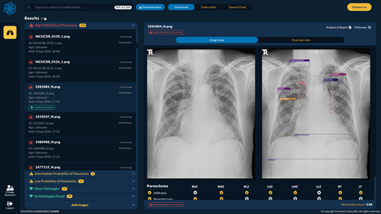
RADIFY® is a radiological computer-aided prioritization, triage, and detection software that utilizes AI-based image analysis algorithms to identify pre-specified findings on frontal chest X-ray images and flag the images in the PACS/workstation to enable prioritized review by the appropriately trained medical specialists who are qualified to interpret chest radiographs. The software does not alter the order or remove cases from the reading queue. It is intended to assist hospital networks and radiologists in workflow triage, identification, and reporting. RADIFY® is therefore providing computer-aided triage (CADt) functionality and computer-aided detection (CADe) functionality
Certification
Envisionit Deep has been awarded ISO 13485:2016 certification by TÜV SÜD. RADIFY® is licensed by the South African Health Products Regulatory Authority (SAHPRA). RADIFY® Triage has received FDA 510(k) clearance.
Development Stage
On the Market
Deployment
Cloud, On-premises, Mobile / Portable, Hybrid
Intended Age Group
The product is intended for patients over six years old, of any gender or race, suspected of having chest abnormalities or pathologies.
Target Setting
The product is intended to be used by private or public primary and tertiary healthcare providers as part of standard clinical workflow, TB screening programs or occupational screening.
Current Market
Southern Africa, United Kingdom, Indonesia
Input
Can be used to read images from any kind of digital X-ray machine and model.
Chest X-ray image format: DICOM, PNG, JPEG.
Chest X-ray type: Posterior-Anterior CXR, Anterior-Posterior CXR.
Output
Output includes:
Heatmap
Dichotomous output indicating whether each abnormality is likely present or absent
Probability score for TB
Probability score for each abnormality
Location of each abnormality
Default threshold for TB is 0.6 and can be adjusted.
A customizable reporting template is available, including ILO standard report, prepopulated based on AI detections.
Lung abnormalities included in the TB Score: Bronchiectasis, Calcification, Cavity, Consolidation, Fibrosis, Lymphadenopathy, Nodule, Opacity, Pleural effusion
Additional findings reported by the product: Atelectasis, Bronchiectasis, Calcification, Cavity, Consolidation, Fibrosis, Lymphadenopathy, Mass, Nodule, Opacity, Pleural effusion, Pneumothorax, Bone Lesion, Cardiomegaly (Cardiothoracic ratio), Cysts, Emphysema, Ground Glass, Peribronchial Thickening, Reticular Opacities, Pneumonia

Hardware
2 CPU cores, 4GB RAM, 1GB HDD, 10mbps network, 1080p high-contrast screen
Server
Intel/AMD x64 platform, 4 CPU cores minimum, 12GB RAM minimum, 150GB system drive. NVIDIA-based GPU is recommended for AI inference acceleraion.
Integration with X-ray Systems
Integration with PACS and Legacy Systems
The RADIFY® Platform allows for the X-Ray images to be received from several integrated sources, such as X-Ray Modality, Hospital PACS system or via RADIFY® Web User Interface or RADIFY® Application Programming Interface (API) services. E.g.
Siemens Healthineers syngo.plaza CI 2019
Yes. RADIFY® fully support DICOM standard. DICOM conformance statement available on request.
Software
Client: Windows 10+ or macOS 12+, compatible web browser: Microsoft Edge 113+, Chrome 114+, Safari 17.5+
Server: Ubuntu 22.04 LTS, Docker or Kubernetes
Processing Time
The RADIFY platform swiftly processes medical imaging data, delivering comprehensive results in just three seconds, enabling rapid diagnostic insights for healthcare professionals.
Data Sharing & Privacy
Data de-identification is available and enabled by default. Details of data sharing and privacy options are available in Envisionit Deep AI® services agreement.
Software Updates
Updates are released every 6 months. Hot fixes and security patches are done based on severity level. Minor versions are backwards compatible with no effect on the users. Major version may include feature deprecation, these are communicated in advance with support from Envisionit Deep AI® staff
Price
RADIFY® is provided with comprehensive pricing options, including volume tiering, specific deployment requirements and level of support required.
Product Development Method
Envisionit Deep AI® uses an Agile development methodology with specific compliance to ISO 13485, ISO14971. IEC62304, and IEC82304.
Training
A geographically diverse dataset in excess of 1.5 million images of paediatric and adult images, including specialist detailed annotations. The dataset is balanced across multiple demographic, image quality, equipment and abnormality subgroups to ensure outstanding generalisation of the AI model.
Reference Standard
Expert human multi-reader annotations, moderated PMS feedback, radiology reports, GeneXpert
Publications
This website works best with browsers other than Internet Explorer.