


Last Updated:
15th May 2023
Chest X-rays are currently the best available method for diagnosing different lung associated diseases like hernia, pneumonia, fibrosis, edema, emphysema, cardiomegaly, pleural thickening, consolidation, pneumothorax, mass, nodule, atelectasis, effusion, infiltrations, and tuberculosis. Our application can detect diagnosis of each of these conditions faster than an average processing time from a radiological laboratory. Especially for Tuberculosis (TB). Our product can be used on a android or IOS phone application or any web browser with a basic WIFI network. It’s easy to use interface makes it an convenient application for any user not well verse with high end technology.
Certification
Certification pending
Development Stage
On the Market
Deployment
Android and IOS app. Our product can be used on a android or IOS phone application or any web browser with a basic WIFI network. It’s easy to use interface makes it an convenient application for any user not well verse with high end technology.
Intended Age Group
0+ years
Target Setting
Primary, public, private
Current Market
India and Africa
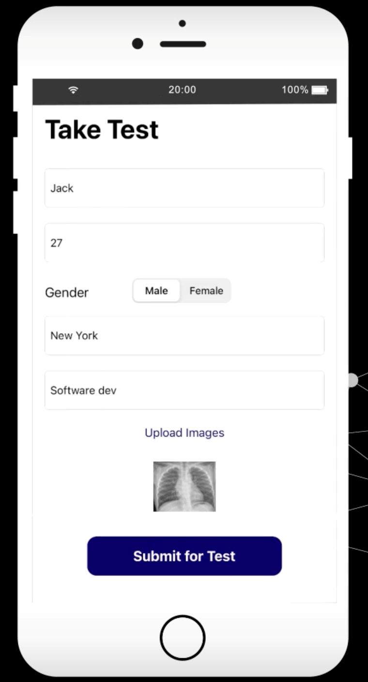
Input
JPG, PNG,
Posterior-anterior CXR
Anterior-posterior CXR
Output
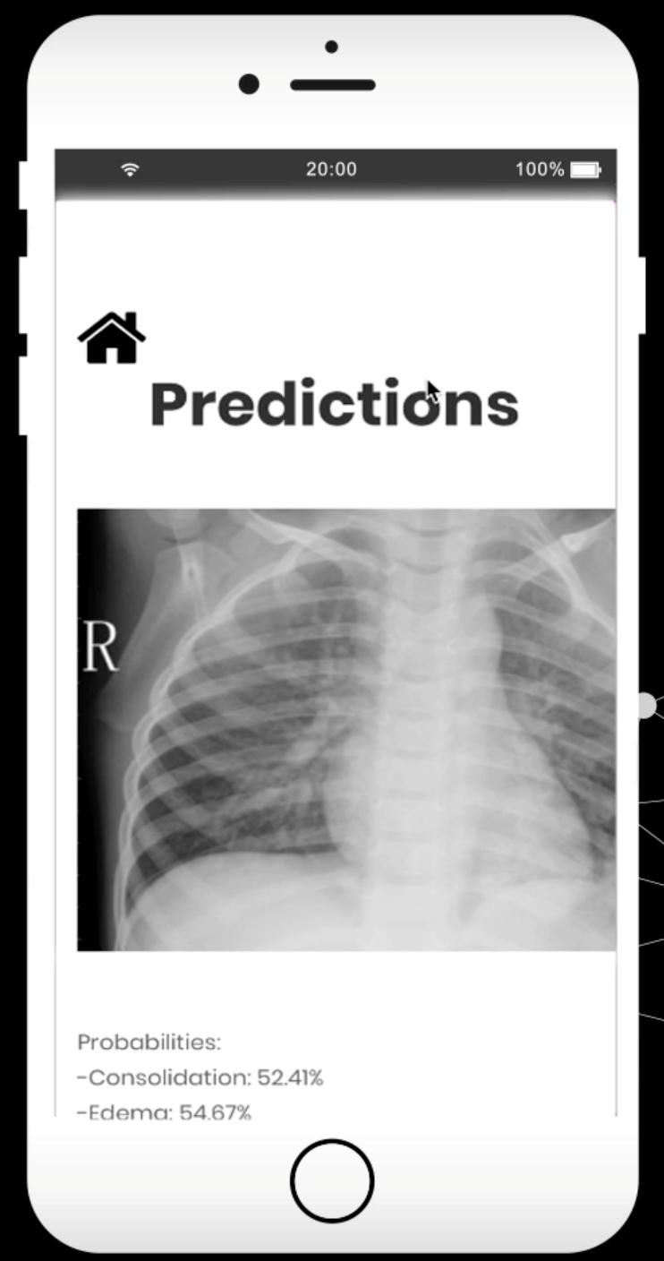
Dichotomous output indicating for each abnormality whether this is present or absent.
Dichotomous output only indicating whether TB is likely present or likely absent.
Probability score for TB
Probability score for each abnormality
Location of each abnormality
The results from ChestAi application are formatted in a structure report to provide information on if disease was detected, if so where was it identified in X-ray image and also information on details of identified condition.
Can detect:
Abscess Air fluid level
Atelectasis √ Blunted costophrenic angle
Calcification
Consolidation Fibrosis
Interstitial markings Loculated pleural effusion
Lymphadenopathy Mass Nodule
Opacity Pleural effusion
Pneumothorax
Hardware
NA
Server
AWS
Integration with X-ray Systems
Integration with PACS and Legacy Systems
The ChestAi application can be integrated in any of the existing hospital data storage PACS platform, the X-ray images normally in JPEG, or PNG formats can be imported into ChestAi to provide diagnosis. If X-ray images are in DICOM format, our platform can convert them to JPEG or PNG format to get it passed through application.
Yes can be integrated
Software
IOS or Android App
Processing Time
1min
Data Sharing & Privacy
None
Software Updates
Software update is scheduled next year Fall 2024 with inclusion of additional use cases including but not limited to hemorrhages, and fractures. No changes will be made to current version of software with existing labels for Tuberculosis and other 16 labels.
Price
Licensing
Product Development Method
The product was developed on java platform, using AWS sagemaker for training, trained models hosted on AWS cloud.
Training
The product was designed and trained on more than 500 thousand X-ray images in total for 16 different conditions.
Reference Standard
The ground truth/reference standard used to diagnose TB condition are patient X-ray with actual TB as diagnosed consistently by more than 5 radiologists in USA. The negative reference are normal X-rays without any condition including TB.
Publications
Single shot detector application for image disease localization, Shrikant Pawar, Aditya Stanam, Rushikesh Chopade, bioRxiv 2021.09.21.461307; DOI: https://doi.org/10.1101/2021.09.21.461307
Neural Networks for Predicting Severity of Ovarian Carcinomas, Rushikesh Chopade, Aditya Stanam, Shrikant Pawar. Intelligent Sustainable Systems. Lecture Notes in Networks and Systems, vol 578. Springer, Singapore. https://doi.org/10.1007/978-981-19-7660-5_7
K-fold Semi-supervised Self-learning Technique for Image Disease Localization, Rushikesh Chopade, Patil Abhijit, Stanam, Shrikant Pawar. Springer Advances in Intelligent Systems and Computing Print ISBN 978-981-19-9818-8. DOI: https://doi.org/10.1007/978-981-19-9819-5_49
This website works best with browsers other than Internet Explorer.